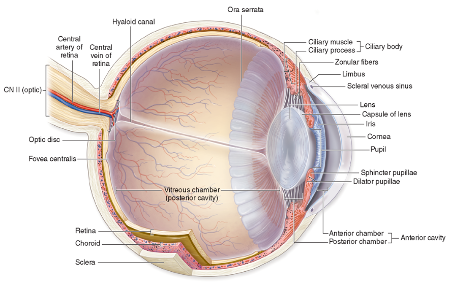
The eye consists of the sclera, uvea, and retina.1
-
Cornea. The sclera is continuous anteriorly as the cornea, which forms a bulging, transparent region specialized for refracting light as it enters the eye. The cornea receives general sensory innervation from the nasociliary nerve (CN V-1).
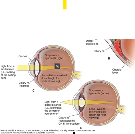
C. Light from a distance is bent by the stretched lens to strike the retina. D. Light from a source nearby is bent even more sharply by the relaxed lens to strike the retina.
The uvea is the middle layer of the eye, sandwiched between the outer sclera (the white of the eye) and the inner retina. It's made up of three main parts:
-
Iris – the colored part of your eye, which controls the size of the pupil and thus how much light enters the eye.
-
Ciliary body – located just behind the iris, this structure helps focus the lens and produces aqueous humor, the fluid in the front part of the eye.
-
Choroid – a layer filled with blood vessels that nourishes the retina and other parts of the eye.
Because the uvea is rich in blood vessels, it’s a common site for inflammation—called uveitis—which can be serious and cause vision loss if not treated.
Let me know if you want to dive into any part more deeply—like diseases of the uvea, or how it's involved in vision.
Content 3
CHOROID
The choroid is the vascular, middle layer of the eye and is composed of the following (Figure 18-2A and B):
The anterior region of the choroid consists of the ciliary muscle, which is composed of smooth muscle and is innervated by parasympathetic neurons from CN III (Figure 18-2B).
-
Ciliary muscle. This circular muscle surrounds the periphery of the lens. Contraction causes the diameter of the muscle to decrease, whereas relaxation causes the diameter of the muscle to increase. Understanding this concept is essential to understanding how the lens focuses on images near and far.
-
Lens. A transparent biconvex structure enclosed in an elastic covering. The lens is held in position by radially arranged suspensory ligaments, which are attached medially to the lens capsule and laterally to the ciliary muscles.
Process by which the lens changes shape to maintain a focused image from distant or near objects.
-
Distant vision. Light rays from distant objects are nearly parallel and do not need as much refraction to bring them into focus (Figure 18-2C). Therefore, to focus on distant objects, the ciliary muscle relaxes, which stretches the suspensory ligaments and flattens the lens. When the lens is flat (least rounded), it is at optimal focal length for distant viewing.
-
Near vision. Light rays from close objects diverge and require more refraction to focus (Figure 18-2D). To focus on near objects, parasympathetic neurons in CN III cause ciliary muscle contraction and thus relaxation of the tension on the suspensory ligaments, allowing the lens to become more rounded. When the lens is round, it is at optimal focal length for near viewing.
The iris is the visible colored part of the eye (Figure 18-2B). The round central opening of the iris is the pupil, which allows light to enter the eye. The iris consists of smooth muscles under autonomic control, which contract reflexively and vary the size of the pupil.
-
Sphincter pupillae muscle. Causes pupil constriction in bright light and close vision via visceral motor (parasympathetic) innervation from CN III.
-
Dilator pupillae muscle. Causes pupil dilation in dim light and distant vision to enable more light to enter the eye via sympathetic innervation.
 Pupillary light reflex. Regulates the intensity of light entering the eye by controlling the diameter of the pupil. Greater intensity of light causes the pupil to become smaller, thus allowing less light to reach the retina. CN II is responsible for the sensory limb of the pupillary reflex by sensing the incoming light. Visceral motor innervation from CN III is responsible for the motor response by constricting the pupil. Light entering one eye should produce constriction of both pupils. An abnormal pupillary light reflex reveals potential problems with CN II, CN III, or the brainstem.▼
The retina is the innermost layer of the eyeball (Figure 18-2A).
-
Optic disc. Consists of the nerve fibers from CN II and thus has no photoreceptors and is insensitive to light, hence the nickname “the blind spot.”
-
Macula lutea. A structure lateral to the optic disc that is specialized for high-acuity vision. Within the macula is the fovea centralis, which contains high concentration of photoreceptors. The eye is positioned such that light rays from the object in the center of the view fall upon this region.
-
Photoreceptors (rods and cones). Rods are specialized cells for vision in dim light (black, gray, white vision), whereas cones are associated with visual acuity and color vision.
The eye is divided into the following segments (Figure 18-2B).
-
Posterior segment. Known as the vitreous chamber, the posterior segment is filled with a clear gel called vitreous humor, which contributes to the intraocular pressure.
-
Anterior segment. Subdivided by the iris into the anterior chamber (between the cornea and iris) and the posterior chamber (between the iris and lens). The anterior segment is filled with aqueous humor, which has a composition similar to plasma. Unlike vitreous humor, which is constant and is never replaced, aqueous humor forms and drains continually into the venous system via the canal of Schlemm.
 Glaucoma. Aqueous humor is produced and drained at a constant rate, and as a result, a constant intraocular pressure is maintained. If drainage of aqueous humor is impaired, the pressure within the eye may increase and cause compression of the retina and damage the optic nerve, resulting in a condition known as glaucoma.▼
The anterior chamber is the space between the cornea and the lens, and it is filled with aqueous humor.
The space between the posterior aspect of the lens and the retina is filled by vitreous gel.
The cornea and the lens form the focusing system of the eye, while the retina functions as the photoreceptor system, translating light to neuronal signals that are in turn transmitted to the brain via the optic nerve and visual pathways. The choroid is a layer of highly vascularized tissue that nourishes the retina and is located between the sclera and the retina. The retinal pigment epithelium (RPE) layer is a monolayer of pigmented cells that are adherent to the overlying retinal photoreceptor cells. RPE plays a major role in retinal photoreceptor metabolism.

e landmarks of the normal fundus. The macula is bounded by the superior and inferior vascular arcades and extends for 5 disc diameters (DD) temporal to the optic disc (optic nerve head). The central part of the macula (fovea) is located 2.5 DD temporal to the optic disc. The peripheral fundus is arbitrarily defined as the area extending anteriorly from the opening of the vortex veins to the ora serrata (the juncture between the retina and ciliary body). (Drawing of Juan R. Garcia from Johns Hopkins University.)
The eyes are photosensitive organs for analyzing the form, intensity, and color of light reflected from objects and providing the sense of sight. They are protected within the orbits of the skull which also contain adipose cushion.
Video: Anatomy of the Eye
External Anatomic Landmarks
Accurate localization of the position of internal structures with reference to the external surface of the globe is important in many surgical procedures. The distance of structures from the limbus as measured externally is less than their actual length. Externally, the ora serrata is situated approximately 5.5 mm from the limbus on the medial side and 7 mm on the temporal side of the globe. This corresponds to the level of insertion of the rectus muscles. Injections into the vitreous cavity through the pars plana should be given 3.5–4.0 mm from the limbus in the phakic eye and 3–3.5 mm from the limbus in the pseudophakic or aphakic eye. The pars plicata, which is the target for cyclodestructive procedures in the treatment of intractable glaucoma, occupies the 2–3 mm directly posterior to the limbus.
The eye is divided into the following segments.
- Posterior segment. Known as the vitreous chamber, it is filled with a clear gel called vitreous humor, which contributes to the intraocular pressure.
- Anterior segment. Subdivided by the iris into the anterior chamber (between the cornea and iris) and the posterior chamber (between the iris and lens). The anterior segment is filled with aqueous humor, which has a composition similar to plasma. Unlike vitreous humor, which is constant and is never replaced, aqueous humor forms and drains continually into the venous system via the canal of Schlemm.
 Aqueous humor normally is produced and drained at a constant rate, and as a result, a constant intraocular pressure is maintained. If drainage of aqueous humor is impaired, the pressure within the eye may increase and cause compression of the retina and damage the optic nerve, resulting in a condition known as glaucoma.
References
1. Orbit. In: Morton DA, Foreman K, Albertine KH. eds. The Big Picture: Gross Anatomy, Medical Course & Step 1 Review, 2nd Edition. McGraw-Hill Education; 2018. Accessed April 11, 2025. https://accessmedicine.mhmedical.com/content.aspx?bookid=2478§ionid=202021018, Chapter 18, Orbit